
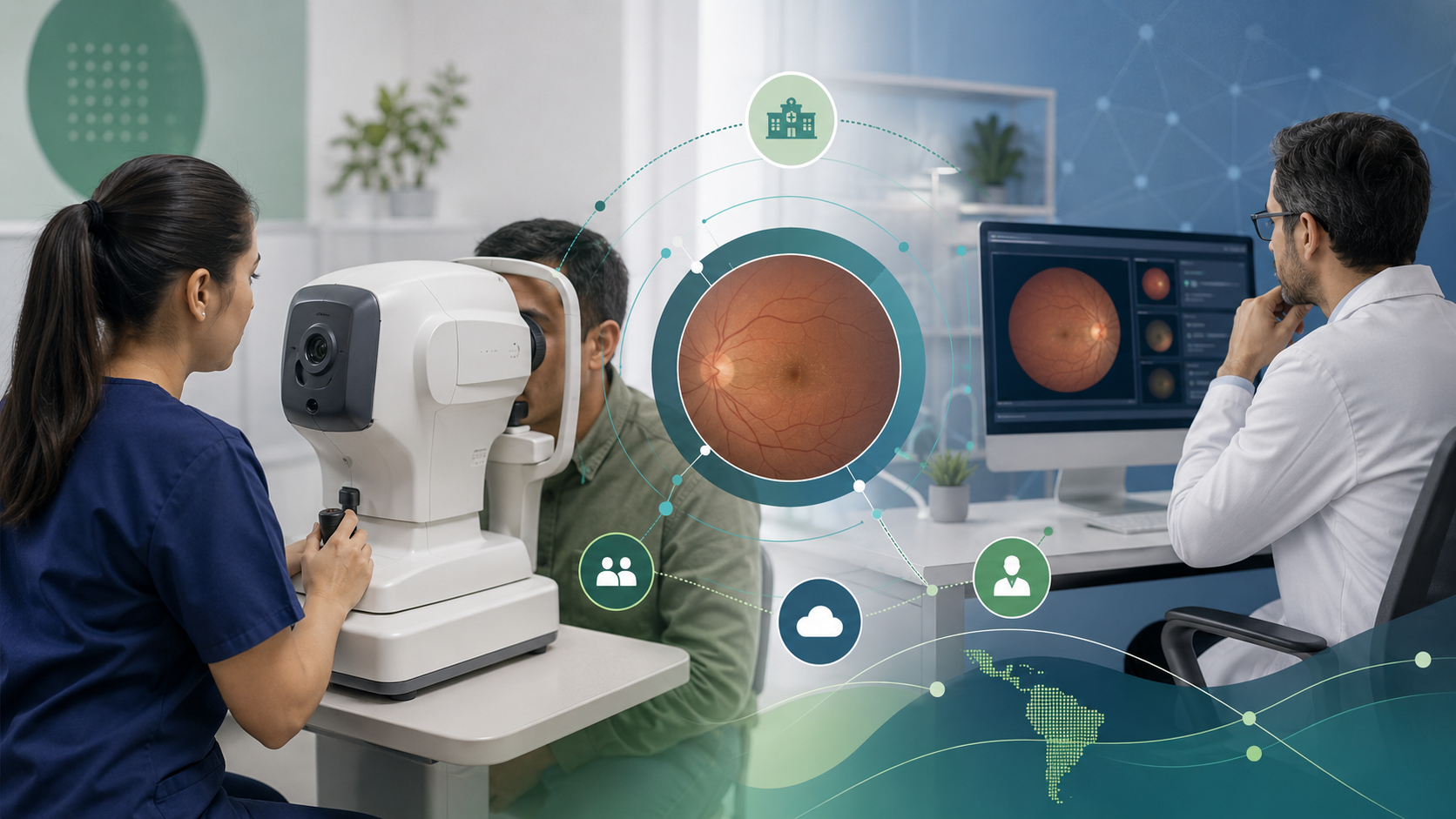
The non-mydriatic fundus camera is often discussed as if it were a complete solution. In reality, it is a valuable component, but still just one component inside a larger workflow.
Its main contribution is operational: it can support retinal image capture with less friction in many care settings and, because of that, it can expand access to eye checks for people with diabetes and other at-risk populations.
What “non-mydriatic” means
In simple terms, it means the device can often capture retinal images without depending on routine pharmacologic dilation in every case. That does not remove all clinical limitations or apply equally in every situation, but it can make logistics easier for screening and periodic control programs.
Why it matters operationally
When the goal is to sustain coverage, every point of friction matters. If a study requires too many steps, more staff, or too much time per patient, the achievable volume drops and adherence suffers.
A non-mydriatic fundus camera can help:
- bring capture closer to non-specialist care settings
- integrate eye checks into diabetes or primary care workflows
- reduce barriers in outreach campaigns or territorial nodes
- make better use of team time
That does not mean every captured image will automatically be useful. Image quality remains critical.
Where it can create the most value
1. Diabetes and chronic care centers
It can add retinal capture without requiring everything to happen inside a traditional ophthalmology service.
2. Hospitals with high demand
It can act as a more organized entry point for screening and prioritization, especially when reading is performed in another hub or through remote support.
3. Territorial networks or outreach campaigns
Its value increases when eye care must be brought closer to populations with access barriers.
What is needed for it to truly transform the workflow
Buying a device is not enough. For it to change operations, institutions should solve several things in parallel:
Image quality
If a large share of studies becomes ungradable, the operational benefit fades. Capture needs training, protocol, and feedback. For more on that, see Retinal image quality: how to reduce ungradable studies in screening programs.
Reading and prioritization
Value appears when capture leads to useful interpretation and an actionable decision. Without that, only the volume of stored images grows.
Referral
A program needs to know what happens to each study: follow-up, recapture, priority review, or referral.
Traceability
Which patient was captured, with what quality, who reviewed the study, and what follow-up was indicated.
Limits worth keeping in mind
A non-mydriatic fundus camera does not automatically solve:
- the need for clinical judgment
- poor capture technique
- the need for human review
- access barriers to specialist care
It also does not make interoperability between devices, software, and records trivial.
How it fits with teleophthalmology
When combined with teleophthalmology, a non-mydriatic fundus camera can enable a more flexible model: capture close to the patient and review where professional capacity exists. That combination makes decentralization more feasible without losing operational order.
How Retinar approaches this
At Retinar, we work with a model designed so teams do not depend on the device alone:
- support for decentralized capture
- focus on real image usability
- risk-based study prioritization
- remote reading and reporting with human review
- integration into the institution’s real operating flow
Conclusion
A non-mydriatic fundus camera can transform diabetes eye care when it is embedded in a workflow for screening, quality, reading, and follow-up. Its greatest value is not the hardware alone, but the ability to bring capture closer to the patient without losing traceability or clinical judgment.
If your institution is evaluating how to use this kind of equipment inside a sustainable operating model, contact us to design the full workflow.